
How does the heart work? Four chambers, two loops and one steady beat
The heart is a muscular pump about the size of a fist that moves blood around the body. Its four chambers work in two loops: the right side sends oxygen-poor blood to the lungs, and the left side pumps oxygen-rich blood to the rest of the body. Valves keep the blood moving one way, and an electrical signal from the heart's own pacemaker sets the beat, which is normally 60 to 100 times a minute at rest.
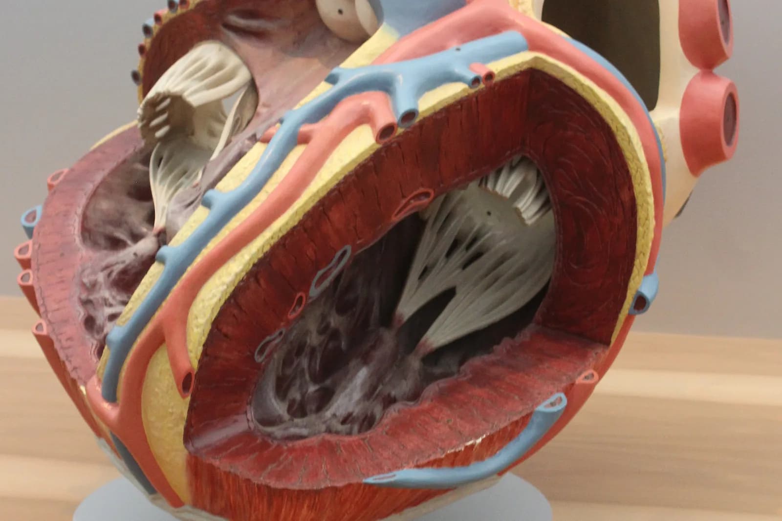
Textbooks describe the heart in a few lines, but the order of events is easier to follow as a story: blood arrives, is squeezed onward, is refreshed in the lungs and is sent out again. This guide follows one beat from the start and explains the lub-dub sound, the two loops and the numbers behind them.
Key ideas
- The heart is about the size of a fist and weighs roughly 250 to 350 grams in an adult.
- It has four chambers, two atria that receive blood and two ventricles that pump it out, with a wall between the sides and four valves.
- Blood travels in two loops: the right side sends it to the lungs, the left side to the body, and the left ventricle pumps hardest.
- Each beat starts in the sinoatrial node; the lub is the valves between atria and ventricles closing, the dub is the exit valves closing.
- At rest the heart beats 60 to 100 times a minute and moves roughly 5 litres of blood a minute.
The pump: four chambers, a wall and four valves
The heart sits in the centre of the chest, between the lungs, tilted slightly to the left. An adult heart weighs about 250 to 350 grams and is about the size of its owner's fist. Its wall is made mostly of cardiac muscle, the myocardium, lined inside by a thin layer, the endocardium, and wrapped in a protective sac, the pericardium.
Inside are four chambers. The two upper ones, the atria, collect blood. The two lower ones, the ventricles, pump it out. A wall of tissue called the septum divides the right side from the left, so the two sides never mix. Four valves act as one-way doors: the tricuspid valve between the right atrium and right ventricle, the mitral valve on the left, and the pulmonary and aortic valves where blood leaves the ventricles.
The heart muscle needs blood of its own. The coronary arteries branch off the aorta and run over the surface of the heart, bringing oxygen-rich blood to the muscle itself.
Two loops: to the lungs and to the body
Blood travels in two connected loops. In the first, oxygen-poor blood from the body enters the right atrium through two large veins, the superior and inferior vena cava. It passes to the right ventricle, which pumps it through the pulmonary artery to the lungs. There the blood takes up oxygen and returns through the pulmonary veins to the left atrium.
In the second loop, the left atrium sends blood to the left ventricle, which builds the high pressure needed to push it through the aorta and the arteries to the whole body. Arteries branch into smaller vessels and finally into capillaries with walls one cell thick, where oxygen and nutrients pass to the tissues and carbon dioxide and waste pass back. Veins then collect the blood and return it to the right atrium.
Textbook drawings colour oxygen-rich blood red and oxygen-poor blood blue. That is a convention only: the blood in your veins is dark red, not blue.
One beat: what lub-dub is
A heartbeat is the contraction of the atria and the ventricles. First the atria squeeze and push blood into the ventricles. A fraction of a second later the ventricles contract and send blood into the pulmonary artery and the aorta. Then the muscle relaxes and the chambers fill again.
The signal for each beat starts in a cluster of pacemaker cells, the sinoatrial node, in the right atrium. It spreads through the atria, pauses briefly at the atrioventricular node so the ventricles can finish filling, and then runs down the walls of the ventricles. The nervous system and hormones speed this rhythm up or slow it down, but the node sets it.
The familiar lub-dub is the sound of valves closing. The lub is the valves between the atria and ventricles shutting once the atria have emptied; the dub is the aortic and pulmonary valves shutting once the ventricles have pumped.
The numbers: pulse, output and a hundred thousand beats a day
At rest, a heart rate of 60 to 100 beats a minute is normal for adults, according to the US National Heart, Lung, and Blood Institute. To find your pulse, place two fingers on the inside of the wrist below the thumb, count the beats for 30 seconds and double the number.
Each beat pushes out a volume called the stroke volume, about 70 millilitres in a resting adult of average size, with a normal range of 55 to 100 millilitres. Multiply the rate by the volume and you get the cardiac output: at 72 beats a minute that is about 5 litres a minute, close to the whole blood volume of an adult. OpenStax gives 5.25 litres a minute as the mean resting output of each ventricle.
At 70 beats a minute the heart beats about 100,000 times a day, since 70 × 60 × 24 is 100,800. Over 80 years that comes to roughly 3 billion beats. During exercise both the rate and the output rise to bring more oxygen to the muscles.
See a heart beat: the VJOURNAL SMART BOOK chapter
The Biology book of the VJOURNAL SMART BOOK series in VITON13 SCHOOL has a chapter called The heart and circulation. A living 3D heart stands beside the text and beats as you read. Its moments follow this guide: the fist-sized pump with four chambers, one beat with its lub-dub, the two circuits drawn as moving dots of blood, the arteries, capillaries and veins, and finally blood pressure and a faster heart.
In the sandbox under the picture you set the heart rate yourself and watch the beat and the output change, then compare the model with your own pulse counted at the wrist. The chapter ends with a short quiz and the list of sources behind its facts, and it is written as a school textbook, not as medical advice.
In everyday language
The heart is a double pump in one case. The right half pushes used blood to the lungs for a fresh load of oxygen, and the left half pushes the fresh blood out to the body. The valves are doors that open only one way.
Try it yourself
Find your pulse with two fingers on the inside of your wrist, count the beats for 30 seconds and double it, then set the same rate on the heart-rate slider in the heart chapter.
Expected result
A resting pulse of about 60 to 100 beats a minute, and a model that beats at the rate you set while showing how much blood it moves.
Check your answer: A heart beats 72 times a minute and pushes out 70 millilitres with each beat. How much blood does it move in one minute?
72 × 70 = 5,040 millilitres, about 5 litres a minute, roughly the whole blood volume of an adult.
Questions
Why does the heart have four chambers?
Two chambers receive blood and two pump it out. Splitting the heart into a right and a left side keeps oxygen-poor blood going to the lungs apart from oxygen-rich blood going to the body, so each loop can work at its own pressure.
What makes the heart beat on its own?
A group of pacemaker cells, the sinoatrial node in the right atrium, produces an electrical signal that spreads through the heart. The nervous system and hormones can speed the rhythm up or slow it down, but the node sets it.
What is a normal resting heart rate?
For adults, 60 to 100 beats a minute at rest is normal, according to the US National Heart, Lung, and Blood Institute. Rates change with activity, and anyone worried about their own heart rate should ask a doctor.
Is blood in the veins blue?
No. Blood is always some shade of red, brighter when it carries a lot of oxygen and darker when it carries less. Veins can look blue through the skin, and diagrams use blue only as a colour code for oxygen-poor blood.
How big is the heart and how much does it weigh?
OpenStax describes a typical heart as about the size of a fist: roughly 12 cm long, 8 cm wide and 6 cm thick. A female heart weighs approximately 250 to 300 grams and a male heart 300 to 350 grams.
Trace one drop of blood: right atrium, right ventricle, lungs, left atrium, left ventricle, body. Then open the heart chapter, press play and compare your own pulse with the model.
Sources and further reading
- NHLBI, US National Institutes of Health: Anatomy of the heart ↗Sources checked:
- NHLBI: How the heart beats ↗Sources checked:
- NHLBI: How blood flows through the heart ↗Sources checked:
- OpenStax Anatomy and Physiology 2e: Heart Anatomy ↗Sources checked:
- OpenStax Anatomy and Physiology 2e: Cardiac Physiology ↗Sources checked: